The Retina
Vision is the primary sense in humans. In our everyday lives, we judge books by their covers, select mates based upon their face structure, shape of their body, skin tone, dress sense, and choose products based upon what's on the outside. It is very difficult for us to see past what we see!
This truth is echoed in our visual system, with a large portion of mammalian brains being dedicated to visual processing. Our primate fovea (the most high-definition part of our retina) is packed with cone shaped photoreceptors which have a one-to-one (though indirect) connection with their respective ganglion cells. This means that, since each ganglion cell is dedicated to only one photoreceptor, much more visual detail can be extracted from the scene. Moreover, about 35 million years ago the primate retina evolved an extra type of colour receptor (the cones), so that while our mammalian cousins tend to be dichromats, we are trichromats. Here is a nice site that gives an example of how it would be to have less photoreceptor types.
On this page I'm going to show a simple little model of the retina. Information that goes to our brain via our eyes undergoes a lot of processing on its way to being recognised as objects (see the Neurons fire & ideas emerge model). However, here I'm just going to focus on the very first layer of this processing - the stuff that happens in the back of the eye.
|
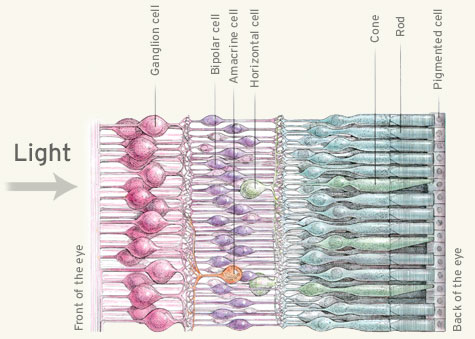
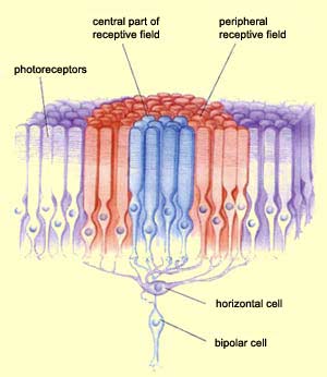
Here's a picture of the different cells in the back of the eye. Light actually travels all the way through all the cells (left to right) before it gets sensed by the photoreceptors, and then travels back the other way (right to left) as neural information.
The cells that we will be modelling here are: the cone photoreceptors, the horizontal cells and the bipolar cells. The output from the retina to the brain is performed by the ganglion cells, but I'm not going to include them in this model because, for our purposes here, their output is not too different from the bipolar cells'. |
|
The problem
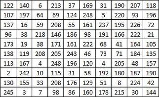
The input to the retina is, obviously, light. It is in the form of a two dimensional array of colour intensities. The problem that the visual system has to solve is the conversion of this 2D array to a symbolic understanding of the objects in the surrounding area. It's difficult for us to grasp the difficulty of this task, since we do it so effortlessly, but if we consider what the photoreceptors are actually sensing, we can see the problem. Here is a 2D array of colour intensities:
|
Can you work out from this what is what objects are in the environment are causing these light intensities?
Our visual system manages this, regardless of many difficulties: the infinite number of different angles that an object can be viewed from, the infinite range of different lighting conditions, the fact that objects may be partially obscured, the distance at which the object is viewed changes the apparent size of the object, the objects are usually moving around, we are usually moving around relative to the objects... |
The problem is solved in stages, and the full process is still far from understood. But the retina's contribution is reasonably well understood, and that's all this model will focus on. The bipolar cells of the retina basically perform two functions: they pick out areas of contrast, like the outlines of objects, and they somewhat reduce the problem of viewing the same object in different lighting conditions. These are the two functional abilities of the retina that we will recreate.
bipolar cells
The bipolar cell is the first cell that shows the famous centre-surround receptive field property. It is by way of this that it picks out areas of contrast and largely ignores differences in lighting.
|
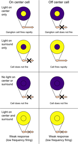
Each bipolar cell receives input from a few photoreceptors in a centre-surround configuration, as shown in the pictures on the right. There are two kinds: ON centre OFF surround cells, and OFF centre ON surround cells. The former is excited by light falling on the centre of its receptive field and inhibited by light falling on the surround of its receptive field. So, maximal excitation is achieved when a light hits all of the centre region and none of the surround, while maximal inhibition is achieved by a ring of light that hits the surround, with a dark patch in the middle. If a light covers both the centre and surround, the excitation of the centre and the inhibition of the surround will more or less cancel each other out, causing either very weak activity or no activity at all.
The OFF centre ON surround cells show the exact opposite behaviour. Now imagine you are looking at the moon in the night sky. All the bipolar cells that are looking at the body of the moon or at the sky will be more or less inactive because their centres and surrounds are uniformly stimulated, so their output will be zero. However, those bipolar cells that are on the circumference of the moon will have uneven light across their centre and surround regions, as shown in the picture below: 
|
The strange conclusion of this is, even though we perceive the moon as a uniform white disc on a black background, what our retina is actually sensing is just the hollow ring that is the moon's circumference. The brain fills in the rest.
|
Horizontal cells
There are two pathways between the photoreceptors and the bipolar cells. The first is the direct pathway, where the photoreceptors that pick up light in the centre of the receptive field are directly connected to the bipolar cell. The other is the indirect pathway, where a horizontal cell intervenes in the connection between the photoreceptors that pick up light in the surround of the receptive field and the bipolar cell. This is how the contrast between centre and surround is achieved.
|
The picture on the right shows the configuration of horizontal cells to a bipolar cell. One bipolar cell is directly connected to the photoreceptors in the centre of its receptive field (the blue cones) and indirectly connected to the photoreceptors in the surround of its receptive field (the red cones) via a horizontal cell. In some cases the horizontal cell may connected to all the cells in the receptive field, whether they be centre or surround. This doesn't make much of a difference because the synapses could be weaker, thus meaning you need more cells to achieve the same result. |
|
testsTo test the model's behaviour, I've taken a couple of tests described in David Hubel's free online book; Eye, Brain, and Vision, plus a general test. I'll describe them below.
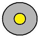
The general test is just to move a bar of light across the receptive field of the bipolar cell. For these examples I'll use the ON centre, OFF surround bipolar cell whose receptive field is shown on the right. The yellow circle is the ON centre, which is excited by light, and the gray ring is the OFF surround, which is inhibited by light. |
|
The general test is shown in the diagram below. The blue bar is a bar of light (the colour is not important). In A1 the light is only hitting a portion of the surround. Since the surround is OFF, this bipolar cell's output will be inhibited so it is less than its resting activity. In A2 a portion of the surround is still being hit by light, but the whole of the centre is covered by the light bar. It is not absolute area that matters, but the proportion of the area, so in the cell in A2, the activation of the ON centre will outweigh the inhibition of the OFF surround, causing this cell to be active above its baseline (resting) level. The case for A3 is fundamentally the same as for A1. In A4 half of the centre and half of the surround are covered by light, so the two will cancel each other out and the cell will remain at the baseline level of activity.
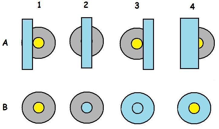
In the lower row, B1 has no light shining on it, so it will be active at its baseline level. B2 has light completely covering its centre, but not touching any of its surround, so it will be maximally active. In B3 the whole receptive field is covered by light, so the centre activation and the surround inhibition will cancel each other out and the overall activity will be the same as in B1 and A4 - baseline level. In B4 the OFF surround is completely covered by light, but none of the ON centre is, so this cell will be maximally inhibited, way below baseline activity.
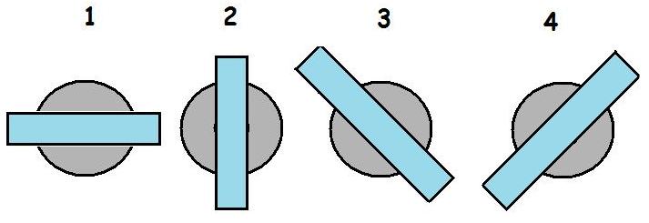
The next test is taken from Hubel's book. Here's the link to where it's described. Basically, if the centre-surround receptive field configuration is correct, then it shouldn't matter what orientation the bar of light is at - if it passes through the centre, then the activation will always be the same. So, in the diagram below, all four cases will cause the same level of activation, because in each case the same proportions of centre and surround are stimulated.
The next test is taken from Hubel's book. Here's the link to where it's described. Basically, if the centre-surround receptive field configuration is correct, then it shouldn't matter what orientation the bar of light is at - if it passes through the centre, then the activation will always be the same. So, in the diagram below, all four cases will cause the same level of activation, because in each case the same proportions of centre and surround are stimulated.
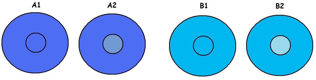
The final test is described here in Hubel's book, and shown in the diagram below. This test shows how it is the contrast, not absolute illumination levels, that the bipolar cells are responsive to. There are two cases below. In case A we apply a uniform light across a cell's receptive field, as in A1. We then increase the brightness of the cell's centre region only, as in A2, and make a note of the increase in activity. The B case is exactly the same, but we start with a brighter uniform light. If the cells were sensitive to the absolute brightness, then B1 should be more active than A1, and B2 should be more active than A2. However, this is not the case. A1 and B1 are active to the same degree (baseline level), and A2 and B2 are active to the same degree.
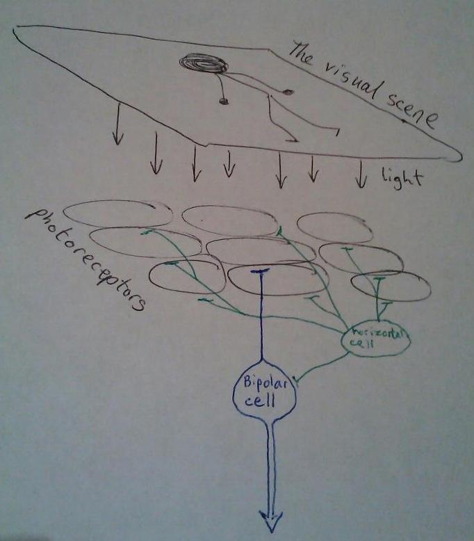
the modelThe model is simple. There will be four layers, three for the retina and one to represent the pattern of light that hits the retina. This is shown on the right.
The top layer is the visual scene (light pattern). Next is a layer of photoreceptors. Then a single horizontal cell. And finally a single bipolar cell. There is a 1 to 1 mapping of light to photoreceptor cells, and since I've chosen to have 9 photoreceptors, that limits the size of the light pattern to 9 units (a 3x3 square). The horizontal cell is responsible for the surround of the receptive field, so it is connected to all photoreceptors except the one in the centre. The bipolar cell is directly connected to the photoreceptor in the centre of its receptive field, and indirectly connected to all the others via the horizontal cell. |
|
In passing, I should mention that it seems that the horizontal cells in the retina are not attached to the cell body of the bipolar cells. Horizontal cells are strange in that they do not have axons, but have dendrites that both receive and send signals. Rather than the horizontal cell --> bipolar cell body configuration shown in my picture, the horizontal cell has a dendrite that intervenes in the synapse between the central photoreceptor and the bipolar cell. Functionally, this is the same as what I have shown here.

|
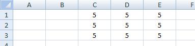
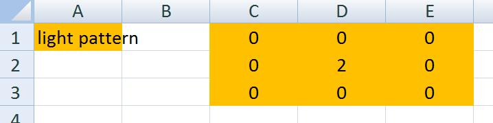
So, for the light pattern, choose a 3x3 block of cells in Excel and put a number 5 in each of them, as shown on the left. I've arbitrarily chosen a brightness scale to be 0-9, so 0 is absolute dark and 9 is the brightest light.
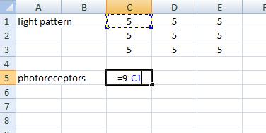
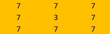
Next, a little bit below our light pattern layer, we will make our photoreceptor layer. Photoreceptors are maximally active in the dark and inhibited by light. To model this, we'll just subtract the corresponding light pattern layer cell''s activity from 9 (our maximum light level). So, click the relevant cell and type =9, a minus sign, and then select the corresponding light pattern cell. Do this for each of the nine cells. On the left is the fully connected photoreceptor layer, all coloured in just because it makes it easier to see :) If you've connected it correctly, when you change the value of any light pattern layer cell, the corresponding photoreceptor layer cell should change to 9 minus that value, as I've shown in the highlighted cell. Have a try and see. Remember: you should only use values of zero to nine. |
You might be wondering why the numbers need to be opposite between the light pattern and the photoreceptors. You're not the only one! However, dark regions are just as useful as light regions in recognition, so we should try to suppress our natural expectation that light be the only thing the retina is looking for. Biology does not have foresight or expectation, so does not work to any plan, and has not concern about what is 'easier' for us to understand - just what functions. For this reason, we'll find many bits that look like they could have been designed better, but just weren't!
|
Next up is the main unit of processing in this model - the bipolar cell. There are two kinds of bipolar cell that we'll look at here, and first of all we'll just do the one that's easier to understand; the ON centre OFF surround bipolar cell.
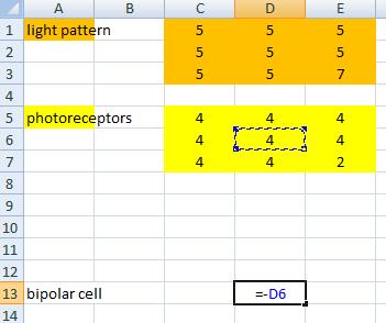
Because we will later have to put a horizontal cell between the photoreceptors and the bipolar cell, choose a cell a little way down the page. The direct pathway connects the centre photoreceptor(s) to the bipolar cell, so the connecting we have to do is very simple. Here's a pic --> Notice the minus sign. An ON centre cell gives positive output to light, so we need to re-reverse the photoreceptors' message (cancel out that minus that we put in the photoreceptor calculation)! |
|
Test that your bipolar cell is working correctly. We will simulate presenting light only to the central photoreceptor by changing just the value of the central cell in the light pattern (D2 on my Excel sheet). If you put a 0 in there, the bipolar cell should output -9, meaning when no light is presented to the photoreceptors, the bipolar cell is maximally inhibited by the photoreceptors. Now try putting 9 into the central light pattern cell. The bipolar cell should output 0, meaning that when maximal light is presented to the centre of the receptive field, the bipolar cell is not inhibited at all by the photoreceptors.
This is all a little hard to follow - photoreceptors being turned off by light, then the bipolar cell being less inhibited as a result. As I said before, this is unfortunate for us, as we have to struggle to get a grip on what's happening, but this is of no concern to the cells in the retina.
This is all a little hard to follow - photoreceptors being turned off by light, then the bipolar cell being less inhibited as a result. As I said before, this is unfortunate for us, as we have to struggle to get a grip on what's happening, but this is of no concern to the cells in the retina.
|
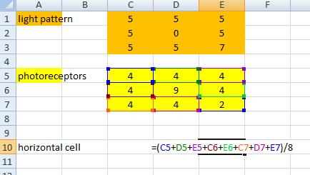
Now it's time to go back and create the indirect pathway from photoreceptors to bipolar cell via a horizontal cell. The horizontal cell is responsible for the surround area of the receptive field, and it does the opposite of whatever the centre is doing. So since the bipolar cell we've made is an ON centre cell, the surround should be OFF. <-- Here's a pic showing how. |
Notice that there's no minus sign, so the inhibition of the photoreceptors is not cancelled out as it was in the bipolar cell. There are 8 surround cells here, and they are supposed to work in roughly balanced opposition to the single centre cell, so I've normalised them by dividing by 8.
|
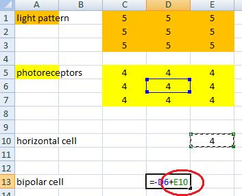
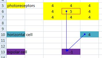
The final step is to complete the indirect pathway by wiring the horizontal cell up to the bipolar cell.
We are making an ON centre OFF surround cell, so the surround connection needs to be opposite in sign to the centre connection. We already connected up the central photoreceptor to the bipolar cell with a minus sign, so the connection from the horizontal cell needs to have a plus sign, like this --> A useful little tool in Excel is the Trace Precedents and Trace Dependents functions in the Formulas tabbed menu. We can see the different inputs to the bipolar and horizontal cells by clicking on one of them and then clicking Trace Precedents. Try it with both --> Once you've done this, have a little play by changing the values in the light pattern layer and observing the resulting bipolar cell activity. |
|
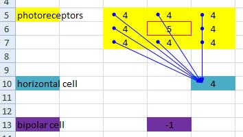
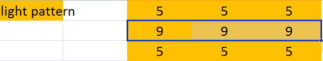
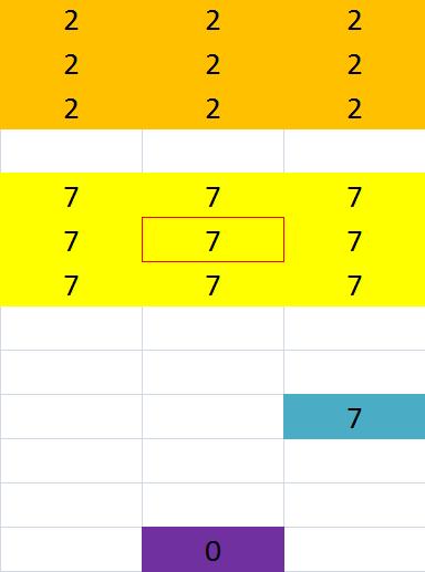
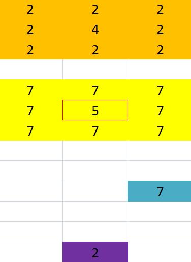
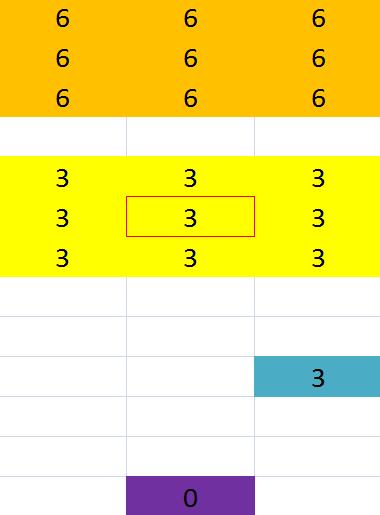
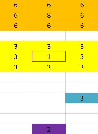
performing the tests1) We set out to make an ON centre OFF surround, cell, so let's start by making sure it works as intended. Set the surround cells to a smaller value than the centre cells, as shown on the right. The centre of the receptive field is brighter (has a higher value) than the surround by 2 units. This stimulus should excite an ON centre OFF surround cell, so as long as the output of the bipolar cell is positive, it's worked. I get a bipolar output of 2, so mine's working.
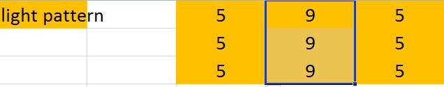
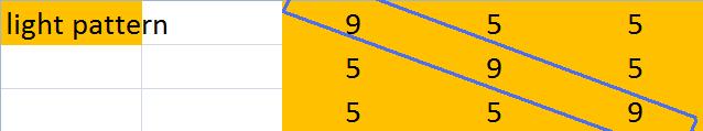
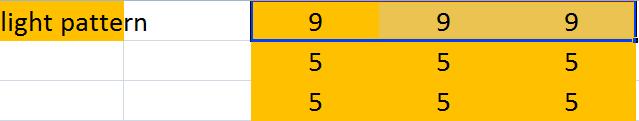
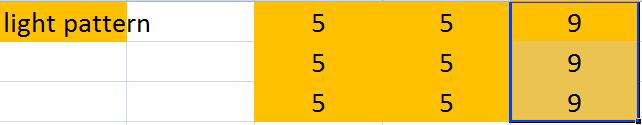
Here are some more inputs you could try --> 2) We also need to make sure that the opposite kind of stimulus inhibits the cell, so try some inputs where the centre of the receptive field is of a smaller light intensity than the surround. Here's an example --> I got a -4 output from the bipolar cell from this input. 3) Now for the contrast test. Remember that the retina doesn't care about absolute brightness, but just brightness differences across its receptive field. So, let's try presenting a uniform brightness of 2 to all cells, then raising the centre cell only to 4 and observing the bipolar cell output. After that, we can try the same thing with a uniform brightness of 6 and raising the centre cell to 8. If our cell is contrast sensitive, the result for both should be exactly the same. This test is shown on the right, with the two uniform brightnesses of different initial intensities shown on the top row. Notice that in each case the bipolar cell is outputting the same value of zero - it is inactive (because it's sensing no contrast). The bottom row shows the two cases where the central cell only has been presented with a light that is two units brighter than the surround. In each case, regardless of the absolute intensity, the bipolar cell is outputting 2. This goes some way to explain how we are able to recognise things in different levels of light without even realising that the light level is different. 4) The final test is to apply bars of light that cross the receptive field at different orientations. On the right there are some example inputs. In the first three cases, the bar crosses the receptive field centre as well as two out of eight of the surround cells. Since, under uniform light, the single centre cell's activation balances with the eight surround cells, when only two of the surround cells are active, the centre has a greater influence than the surround and the bipolar cell becomes excited (with a value of 3 in each of the cases shown here). In the next two pictures, I've shown bars of light that hit just the surround without touching the centre. A light on the surround of an OFF surround cell causes inhibition in that cell, so the bipolar cell will output a negative number. -1.5 in these two cases. |

|


off centre bipolar cells
The model above is only of an ON centre OFF surround cell. But I said earlier that bipolar cells come in two flavours, the other one being the OFF centre ON surround kind.
After making the ON centre cell above, you should be able to make an OFF centre cell yourself just by reversing the signs from the photoreceptors to the bipolar cell in the direct pathway, and from the horizontal cell to the bipolar cell in the indirect pathway. Try this yourself.
In case you get confused, or are just too lazy, here is the one I made:
| retina.xlsx |
